Electron microscopy
At Paihau—Robinson Research, electron microscopy is much more than a technical capability—it is a window into the unseen structures that shape our world.
Unlocking the microscopic universe—electron microscopy for science, industry, and education
Our state-of-the-art scanning electron microscopy facilities serve a broad community, from advanced materials scientists to secondary schools and industry seeking practical answers. Through our suite of advanced instruments, we help customers and collaborators visualise and analyse materials at resolutions far beyond what traditional light microscopes can offer.
Whether you are an academic researcher, a business in need of advanced materials analysis, or an educator eager to inspire your students, the Paihau—Robinson Research Institute’s electron microscopy services offer expertise, access, and a passion for discovery. Contact us today to discuss how our facilities can help you see more, understand more, and do more.
Our state-of-the-art equipment
Our advanced microscopy facilities are anchored by a suite of cutting-edge electron microscopes, supporting research, industry, and education across Aotearoa New Zealand.
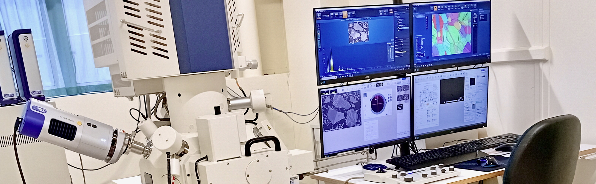
Our laboratory-based Hitachi SU3800 Scanning Electron Microscope (SEM) is the core of our analytical capability, offering rapid imaging and elemental analysis through Energy Dispersive X-ray Spectroscopy (EDS), techniques vital for metallurgical studies, quality assurance, and innovative materials research. Complementing this, the Hitachi SU7000 Field Emission SEM takes imaging and elemental analysis to the next level. This high-resolution instrument enables imaging at a nanoscale and provides additional hardware and software capabilities in elemental analyses, supporting both industry and academic researchers explore the frontiers of modern science.
Our commitment extends beyond the lab with the TM4000Plus portable SEM, empowering our outreach programme to bring real-world science experiences to schools across the country. This mobile, user-friendly microscope allows students and teachers to examine the micro and nanostructures of everyday and advanced materials first-hand, inspiring the next generation of scientists and engineers.
Learn more about each instrument and how they can support your research, industry needs, or educational goals.

Hitachi SU7000
For those seeking to explore materials at the frontier of modern science, the Hitachi SU7000 Field Emission SEM provides high-resolution nanoscale imaging.

Hitachi SU3800
Our flagship Hitachi SU3800 Scanning Electron Microscope (SEM), forms the heart of our analytical services.

Portable TM4000Plus
Through our science outreach programme, we bring cutting-edge technology directly to New Zealand schools.
Consistently great feedback from our customers
Our commitment to quality and client support is reflected in the words of our customers.
Having used Victoria University's SEM facilities for many years, I cannot recommend them highly enough. Sarah is absolutely fantastic to deal with and does her utmost to process our specimens as soon as humanly possible. Results are always provided promptly and any queries are quickly responded to.
Metallurgical and Industrial Consultants, Wellington
Robinson Research provided our company with very valuable, prompt and accurate SEM services, which enabled us to make well-founded decisions on our Floeting® Diamond innovation, further strengthening its position in the market
The Village Goldsmith, Wellington
Sarah is super friendly and welcoming to talk to and chat about the SEM and its capabilities. The imaging and elemental analysis are easy to navigate and switch between. Would definitely recommend their services, especially for first-time users who do not know much about SEM.
Asure Quality, Forensics Division, Wellington
Avertana relies heavily on Sarah to image and analyse samples, both for product quality and for mineralogy studies of slags and minerals. These include high magnification SEM imaging as well as lower magnification EDX studies. She runs an efficient and reliable service, which we would recommend.
Avertana, Auckland
The people
Sarah Spencer has more than 20 years of experience in electron microscopy and runs the scanning electron microscope facility.
Scientist
Robinson Research Institute