Hitachi SU3800
Our flagship Hitachi SU3800 Scanning Electron Microscope (SEM), forms the heart of our analytical services.
Our flagship Hitachi SU3800 Scanning Electron Microscope (SEM) forms the heart of our analytical services. Together with its integrated Oxford Energy Dispersive X-ray Spectroscopy (EDS) detector, this robust instrument enables secondary electron and backscatter imaging alongside quick and accurate elemental analyses on a diverse range of materials.
Non-conductive samples from both biological and man-made sources, such as plants, insects, textiles, rubber, and plastics, can be observed using the variable pressure feature. This enables sample observation without the need for any additional sample preparation, facilitating fast turnaround.
Under traditional SEM conditions, a vast array of sample types can be examined. Examples include analysis of corrosion in metal, steel characterisation, properties of coatings on paints, and mineral identification in geological materials. Such work can be carried out by observing the surfaces of material or by using sample preparation techniques available in our facility, enabling the inner structures of a material to be exposed, which provides an even greater understanding of a material or process.
The instrument is also user-friendly, with local customers having the option of being trained to perform their own work at a reduced cost. The versatility makes the SU3800 an indispensable tool, empowering organisations to innovate, solve problems, and maintain excellence in their respective fields.
SU3800 sample images

Flower
Secondary electron image of a flower using variable pressure mode, eliminating the need for a conductive coating.
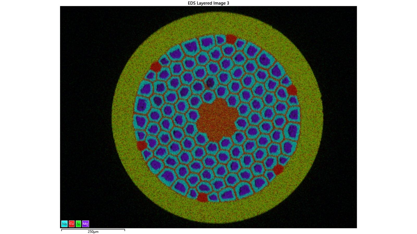
Wire
EDS map of a polished cross-section of superconductor wire.

Ilmenite
Backscatter image of an ilmenite particle showing Si-rich inclusions (darker regions).